
Every day that goes by and we don’t win a ballgame, that’s a missed opportunity for us to battle back. —Brett Gardner
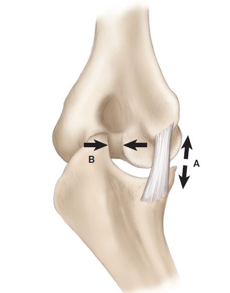
First, hard throwers, including pitchers and position players, not only impose great force on their UCL, they also put great force on the bone at the back of the elbow, called the olecranon (Figure 1). This additional stress can create bone spurs, also called osteophytes. Similar to how stress on skin, such as to your hand with batting, can cause skin callus. The bone spurs can crack and become loose, which is often referred to as “chips in the elbow” (Medically, bone chips are referred to as loose bodies). When the bone spurs break, they can cause significant pain in the back of the elbow. Because the location of pain is near the triceps tendon, many players incorrectly believe they have triceps tendinitis.
What does it mean if you have a bone spur or a loose chip?
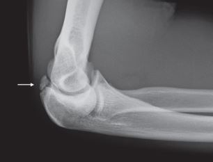
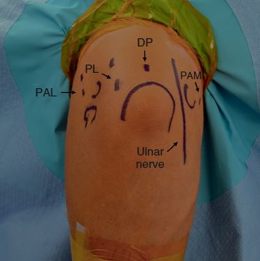
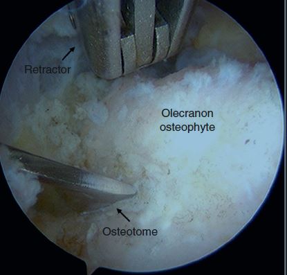
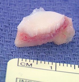
It is absolutely necessary to make an accurate diagnosis, start by knowing what to look for. Figure 2 shows example x-rays of a bone chip. Once properly diagnosed, treatment will depend on the severity of the symptoms. Some players can get through the season with anti-inflammatory medication, and even a cortisone injection. Other players will experience pain to a debilitating degree, and require surgery. Fortunately, bone chips can be removed arthroscopically, and as you can see in figures 3, 4, and 5 and the pain related to the bone chips is eliminated.
We’ve observed a relationship between bone chips that form in a thrower’s arm, and injury to the UCL. For some players, an injury to the UCL may have occurred, and the slightly stretched ligament will result in the olecranon bone having to absorb compounding stress with each throw. I was able to prove this during a research study over 10 years ago, and leverage that knowledge today to perform careful examinations of every player’s UCL who has a bone chip in their elbow. Not every examination requires an MRI, but it’s important to know when you absolutely do.